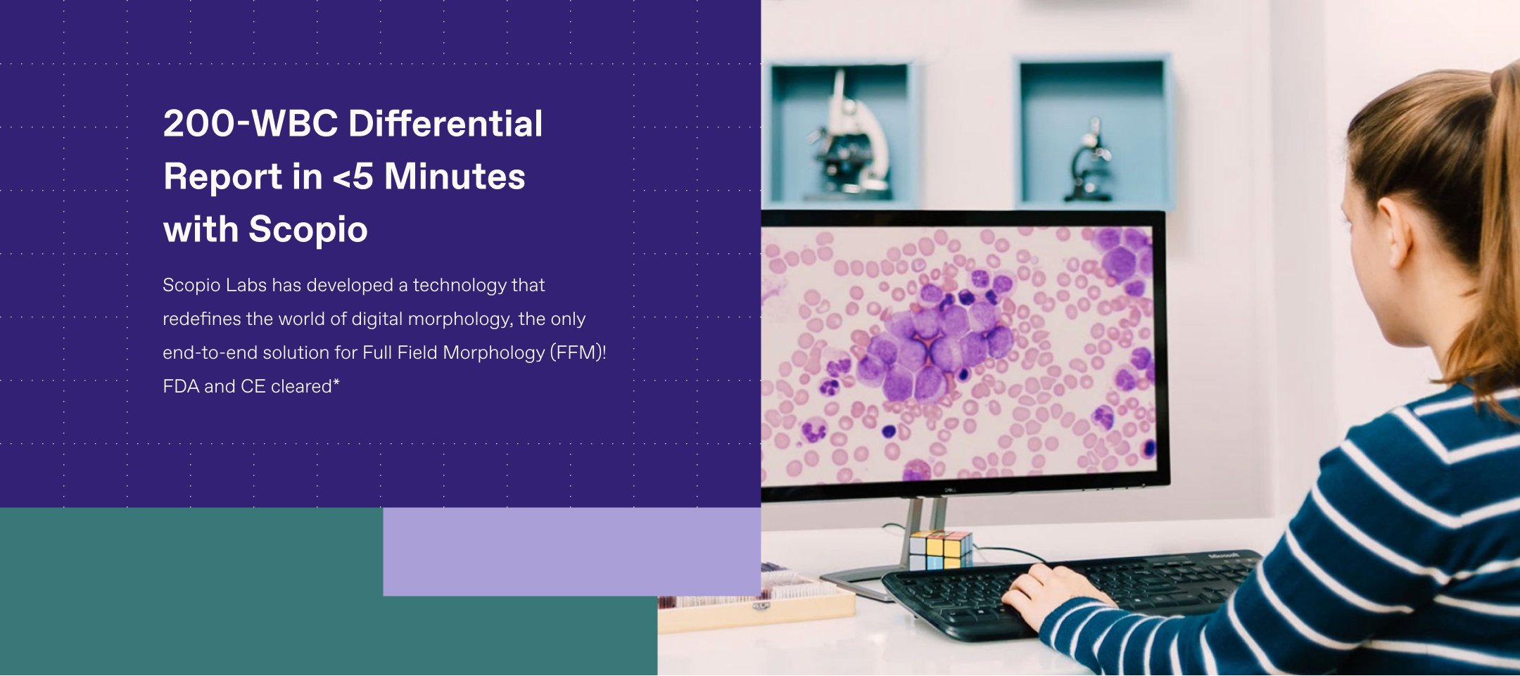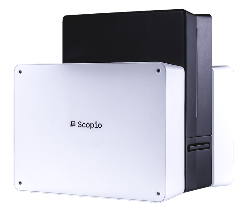
Scopio Labs has developed a technology that redefines the world of digital morphology, the only end-to-end solution for Full Field Morphology (FFM)!
FDA and CE cleared*


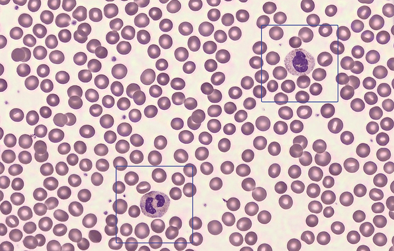
We believe in focusing on the small details, without losing the big picture. While other microscopy-solutions offer only single-cell snapshots, our FFM technology provides cell images at X-100 magnification, with a full field view large scan of the monolayer and the feathered edge area.
Finally, you get the real impression and full context of the slide just like you would in your manual microscope, without leaving your computer’s screen!
Available now for Full Field Morphology Peripheral Blood Smear (FFM-PBS) application. Bone Marrow Aspirate (FFM-BMA) application is under clinical studies (commercial launch expected in 2021).

FFM-PBS scan taken with the X-100 by Scopio Labs


| Scopio X-100 |
Other solutions | |
|---|---|---|
| Technology | Full Field Morphology (FFM) | Snapshots of Single Cells |
| Slide Throughput | 3 Slides Tray | 1 Slide Tray |
| Up to 15 Slides/h for 200** WBCs Differential | Up to 10 Slides/h for 100 WBCs Differential | |
| Adaptive Monolayer Detection |  Supporting Long and Short Smears Supporting Long and Short Smears |
 |
| Feathered Edge Impression |  Included in the Scan Included in the Scan |
 |
| Number of WBC Analyzed | Up to 200** (with no effect on scan time) |
100 |
| Number of WBC Classes | 16 | 12 |
| RBC Analysis Area | 1000 FOVs | 9 FOVs |
| Scanned Monolayer Area | 1000 FOVs | 9 FOVs |
| Platelet Pre-Estimation |  From 10 High Power FOVs From 10 High Power FOVs |
 |
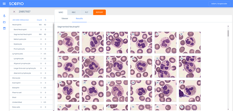
One central element of our end-to-end solution is the Artificial Intelligence (AI) tool, designed explicitly for high-res microscopy and was validated for accuracy and specificity in clinical studies.
Another element is the remote connectivity feature that enables digital-sharing of scanned slides for remote consultation and report generation, without the need to send the slides physically!
Combining these two features with our FFM technology will optimize your lab workflow and substantially reduce diagnosis time cycles. This can improve the consistency of results while maintaining high standards of your decisions.
If your hematology lab is practicing manual workflow and considering a semi-digital solution, you can now instead take the digital leap and use our full-field solution.
Enter your contact details and our team will be in touch